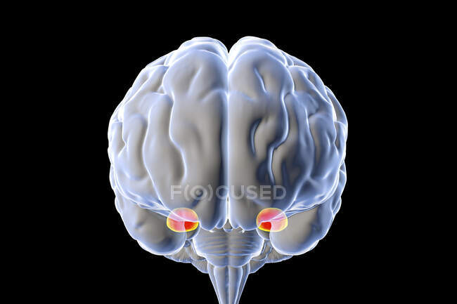
The hypothalamus is a small, central region of the human brain formed by nervous fibers and a conglomerate of nuclear bodies with various functions. Īn important role in hypothalamic development is assigned also to the presence of specific signaling centers (Wingless-Int protein family–Wnt, Hedgehogs family–Hh, and Bone morphogenetic family–FgF) that modulates cell proliferation and neurulation. This condition puts the diencephalon rostrally between the telencephalon cranially and the midbrain caudally and sets the hypothalamus independent from the diencephalon as a distinct posterior part of the forebrain.

According to Puelles’ Prosomeric model, the initially proposed longitudinal axis of the brain is bent due to the first mesencephalic flexure of the embryo. In the last decades, mapping of the genes involved in hypothalamic development allowed the identification of a disparity between the morphological, classic boundaries of this region and the molecular ones. The columnar morphologic model is based on the division of the forebrain in functional longitudinal units, placing the telencephalon in the most rostral region and the diencephalon caudally, in between the telencephalon and the midbrain, while the hypothalamus if formed from the ventral most part of the diencephalic vesicle. Since Herrick first proposed the columnar model of the forebrain organization, the anatomical description was accepted per seand very few research papers have questioned its validity. The hindbrain vesicle or rhombencephalon divides in metencephalon, which further forms the pons and the cerebellum and the myelencephalon that forms the medulla.Įmbryological concepts regarding the development of the hypothalamic region are over 100 years old. Mesencephalon forms the midbrain, structure involved in the processes of vision and hearing. Prosencephalon further divides into two secondary vesicles, the telencephalon that will form the cerebral hemispheres and the diencephalon which gives rise to the diencephalon. Embryological development of the hypothalamusĪt the end of the fourth week of embryological development, the neural tube is organized in primary vesicles: the forebrain vesicle or prosencephalon, the midbrain vesicle or mesencephalon, and the hindbrain vesicle, also called rhombencephalon.


 0 kommentar(er)
